Shop No. 19, Meera
Arcade, Plot No. 14,
Sector-20, Kharghar, Navi Mumbai 410210
Sonosalpingography (SSG)
Sonosalpingography
Sonosalpingography (SSG), also known as saline infusion sonography (SIS), is a specialized ultrasound procedure used to examine the uterus and the uterine lining. It is commonly employed to investigate abnormal uterine bleeding, infertility, recurrent miscarriages, or to evaluate the uterine cavity before in vitro fertilization (IVF) procedures. SSG can detect abnormalities such as polyps, fibroids, adhesions, and congenital anomalies within the uterine cavity.
SSG is a minimally invasive test that helps physicians diagnose conditions affecting the uterus and assists in planning appropriate treatments. It is often performed when standard transvaginal ultrasound results are inconclusive or when more detailed visualization of the uterine cavity is needed.
Benefits & Risks
There are several benefits of performing a sonohysterography test compared to other diagnostic methods:
- SSG is minimally invasive and typically performed on an outpatient basis.
- It provides detailed images of the uterine cavity and can detect abnormalities not visible on standard ultrasound.
- The procedure helps in identifying blockages or abnormalities that may not be detected by ultrasound or other imaging techniques.
- It can assist in diagnosing the causes of infertility and guide subsequent treatments.
Although, there are no major risks associated with SSG, some of them may be :
- Mild to moderate discomfort or cramping may occur during and after the procedure.
- There is a slight risk of infection, which can be minimized by following pre-procedure instructions and using sterile techniques.
- Rarely, an allergic reaction to the saline solution used during the procedure may occur.
How it is performed ?
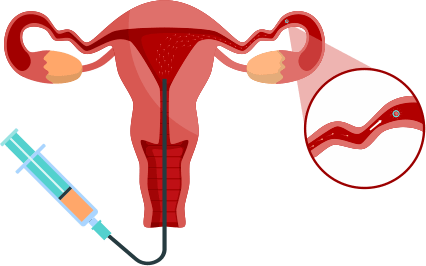
During sonosalpingography (SSG), the patient lies on an examination table, and a speculum is inserted into the vagina to access the cervix, which is then cleaned with an antiseptic solution. A thin catheter is gently inserted through the cervix into the uterine cavity, and sterile saline solution is infused to distend the cavity and fallopian tubes. A transvaginal ultrasound probe is then used to capture detailed images of the uterine cavity and fallopian tubes, highlighting any blockages or abnormalities. The entire process typically takes about 15-30 minutes and is minimally invasive, with patients experiencing mild discomfort or cramping that generally resolves quickly.
Where it is used ?
Sonosalpingography (SSG) is used in specialized fertility clinics, hospital radiology departments, and outpatient imaging centers to diagnose conditions related to infertility, recurrent miscarriages, and to evaluate the uterine cavity and fallopian tubes before in vitro fertilization (IVF) procedures.
.png)
.png)
.png)