Shop No. 19, Meera
Arcade, Plot No. 14,
Sector-20, Kharghar, Navi Mumbai 410210
Color Doppler
Color Doppler Test
Color Doppler ultrasound is a non-invasive diagnostic imaging technique that combines conventional ultrasound with Doppler ultrasound. It is used to visualize blood flow within the body's blood vessels and organs, providing valuable information about circulation and detecting abnormalities.
How Color Doppler Works
Color Doppler ultrasound uses high-frequency sound waves to create real-time images of blood flow. The Doppler effect is utilized to measure the speed and direction of blood flow within vessels, which is represented on the ultrasound screen in color-coded patterns. Red indicates blood flow towards the ultrasound probe (positive Doppler shift), while blue indicates blood flow away from the probe (negative Doppler shift).
Clinical Applications of Color Doppler
Vascular Imaging:
Color Doppler is used to evaluate blood flow in arteries and veins throughout the body, helping diagnose conditions such as peripheral artery disease (PAD), deep vein thrombosis (DVT), and carotid artery disease. It provides detailed images of vessel morphology, detects stenosis (narrowing), and assesses the effectiveness of treatments such as angioplasty and stenting.
Cardiac Imaging:
In cardiology, color Doppler assesses blood flow through the heart chambers and valves, aiding in the diagnosis of congenital heart defects, valve abnormalities (e.g., regurgitation or stenosis), and assessing heart function. It helps cardiologists evaluate the severity of conditions like mitral valve prolapse or aortic aneurysms.
Obstetrics and Gynecology:
Color Doppler is used in obstetrics to monitor fetal blood flow in the umbilical cord and placenta, ensuring adequate oxygenation and growth. In gynecology, it assesses blood flow to ovarian masses, uterine fibroids, and endometrial abnormalities, aiding in the diagnosis of conditions like ovarian torsion or endometrial polyps.
Organ and Tissue Evaluation:
Color Doppler evaluates blood flow within organs such as the liver, kidneys, and spleen, detecting abnormalities such as tumors, abscesses, or cysts. It assists in guiding biopsies and assessing organ perfusion in cases of trauma or transplant evaluation.
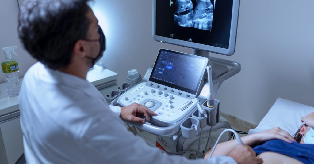
Conducting the Color Doppler Test
Color Doppler ultrasound is performed by trained sonographers or radiologists in hospital settings, outpatient imaging centers, or specialized clinics. A water-based gel is applied to the skin over the area of interest, and a handheld transducer is moved gently to capture images and Doppler signals. Real-time images and Doppler waveforms are displayed on a monitor, and the entire procedure is painless and typically takes 30-60 minutes, depending on the area being examined.
Color Doppler ultrasound is a versatile imaging modality that enhances diagnostic accuracy by providing detailed information about blood flow dynamics and vascular anatomy. It plays a crucial role in various medical specialties, aiding in the early detection, diagnosis, and management of a wide range of vascular and organ-related conditions.
.png)
.png)
.png)