Shop No. 19, Meera
Arcade, Plot No. 14,
Sector-20, Kharghar, Navi Mumbai 410210
Ultrasonography
Ultrasonography
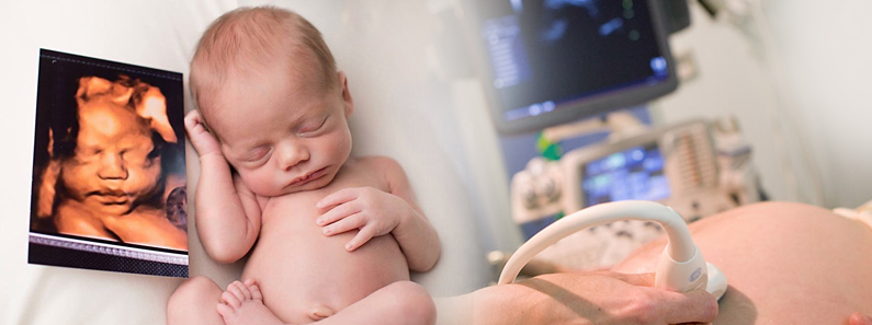
Ultrasound imaging uses sound waves to produce pictures of the inside of the body. It is used to help diagnose the causes of pain, swelling and infection in the body’s internal organs and to examine a baby in pregnant women and the brain and hips in infants. It also used to help determine and perform biopsies, heart conditions, and damage after a heart attack.
Ultrasound imaging is a noninvasive medical test which will help physicians diagnose conditions and help them treat it effortlessly. Ultrasound is used to help physicians evaluate symptoms such as, pain, swelling and infection.
Benefits & Risks
There are many benefits of performing an ultrasound over an MRI or any other non-invasive procedures :
- ultrasound scanning is noninvasive and painless.
- ultrasound is widely available, easy-to-use and less expensive than other imaging methods.
- ultrasound imaging is extremely safe and does not use any ionizing radiation.
- ultrasound scanning gives a clear picture of soft tissues that do not show up well on x-ray images.
Although, there are no major risks associated with ultrasound imaging, some of them may be :
- ultrasound exams in which the transducer is inserted into an opening of the body may produce minimal discomfort.
- most ultrasound examinations are completed within 30 minutes, although more extensive exams may take up to an hour, when the examination is complete, you may be asked to dress and wait while the ultrasound images are reviewed.
How it is performed ?
Ultrasound is safe and painless, and produces pictures of the inside of the body using sound waves. Ultrasound imaging, or sonography, involves the use of a small transducer and ultrasound gel placed directly on the skin.
High-frequency sound waves are transmitted from the probe through the gel into the body. The transducer collects the sounds that bounce back and a computer then uses those sound waves to create an image. Ultrasound examinations does not expose the patient to any radiation.
Where it is used ?
Ultrasound is a safe, fast and efficient way of examining many of the body parts inside a patient which include, heart and blood vessels, liver, gallbladder, spleen, pancreas, kidneys, bladder, uterus, ovaries, eyes, thyroid and parathyroid glands, scrotum, brain, hip and spleen in infants.
Ultrasound is preferred imaging modality for the diagnosis and monitoring of pregnant women and their unborn babies, ultrasound provides real-time imaging, making it a good tool for guiding minimally invasive procedures such as needle biopsies and fluid aspiration.
.png)
.png)
.png)